


15 Oct What I Look for Before Treating Hyperpigmentation: Doctor’s PoV
What I Look for Before Treating Hyperpigmentation
By Dr Rachel Ho | Updated September 2026
Most patients who see me about pigmentation have already tried numerous brightening creams, chemical peels or different laser treatments. Sometimes, the results of creams and treatments are not satisfactory; with incomplete lightening or recurrences. In my opinion, recurrences or recalcitrant hyperpigmentation is not a failure of treatment, but rather, they reflect the complexity of treating hyperpigmentation.
A typical patient from Singapore in my clinic has more than a single type of hyperpigmentation, and the hyperpigmentation can reside in different depths of the skin. Before embarking on laser or any pigmentation lightening treatment for my patients, this is how I assess each patient for their diagnosis, safety and suitability.
Why diagnosis of hyperpigmentation is necessary before starting laser treatments
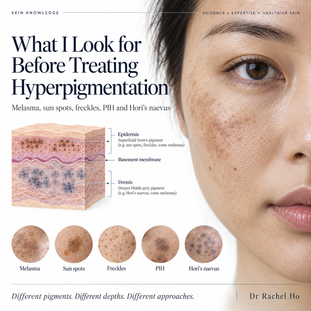
The terms “dark spots” and “hyperpigmentation” are often loosely used to describe darker patches on the skin due to excess melanin deposition. However, hyperpigmentation is a description or symptom, not a diagnosis.
The following patterns of hyperpigmentation can depth of melanin shapes the diagnosis and treatment for patients:
- Depth: Epidermal pigment sits near the surface and tends to clear more easily and faster than deeper melanin deposition. Dermal pigment is located deeper, is often out of reach to topical creams and requires multi-modality treatments and a longer course.
- Behaviour: Some types of hyperpigmentation are stable, but diagnoses like melasma can be reactive and worsened by heat, light, hormones and inflammation.
- Safety: A dark spot that is rapidly changing in size, shape or colour needs to be evaluated first to exclude malignancy before embarking on pigmentation lightening treatments¹.
It is common for patients to present with more than a single type of hyperpigmentation. For example, I often notice melasma, solar lentigines and post inflammatory hyperpigmentation from acne in the same patient.
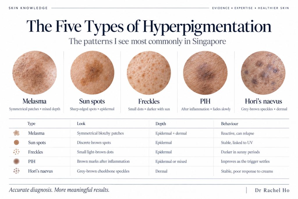
Melasma, sun spots, freckles, post inflammatory hyperpigmentation and Hori’s naevus differ in their appearance, pigment depth and behaviour. This comparison summarises the five patterns discussed in Dr Rachel Ho’s hyperpigmentation guide.
The five types of hyperpigmentation I see most commonly in Singapore
Melasma
Melasma tends to present as symmetrical brown or grey-brown patches with soft edges, on both sides of the cheeks, forehead, nose or upper lip. Melasma darkens with sun, visible light, heat and hormonal changes such as pregnancy, contraceptive, perimenopause and hormone replacement therapy. Microscopically, the melanin deposition occurs in the epidermis and the dermis²,³ .
Sun spots (solar lentigines)
Also known as age spots and liver spots, solar lentigines are brown spots on sun-exposed skin such as the face and backs of the hands with well defined margins. They build up with cumulative UV exposure and become more obvious with age. Pigmentation typically is located in the epidermis.
Freckles (ephelides)
Freckles are little light-brown dots that appear from childhood, mostly across the nose and cheeks. They darken after sun exposure and fade in low-sun periods. There is a familial predisposition for freckles, and the pigmentation is in the epidermal layer.
Post-inflammatory hyperpigmentation (PIH)
Post-inflammatory hyperpigmentation refers to the brown marks left after inflammation or injury such as acne, eczema, insect bites, burns, harsh products or procedures. For patients with darker skin, the risk of PIH injury or inflammation is higher and can last for a longer duration.
PIH can occur in the epidermis and/or dermis. Brown, epidermal PIH fades faster and responds better to treatment than grey-brown, dermal PIH5. In treating patients with PIH, I often recommend prioritising the trigger first to minimise recurrences of PIH.
Hori’s naevus
Hori’s naevus refers to the grey, slate or blue-brown speckles over both cheekbones, especially in females of East Asian descent. Hori’s nevus usually appears in adulthood. The pigmentation in Hori’s naevus is located deeper in the dermis, so Hori’s naevus does not respond to topical medications or creams. Staged laser treatments are often required to remove Hori’s naevus.
Common types of hyperpigmentation in Singapore
| Type of hyperpigmentation | Appearance | Depth of hyperpigmentation | Behaviour of pigmentation |
|---|---|---|---|
| Melasma | Symmetrical patches, soft edges | Epidermis and dermis | Darkens with sun, heat and hormones; relapses |
| Sun spots | Single, sharp-edged brown spots | Epidermis | Stable; new spots form with sun exposure |
| Freckles | Small light-brown dots | Epidermis | Darker in sunny periods |
| PIH | Brown marks at sites of past inflammation | Mostly epidermis | Fades slowly once the trigger stops |
| Hori's naevus | Grey-brown speckles on both cheekbones | Dermis | Stable; does not respond to creams |
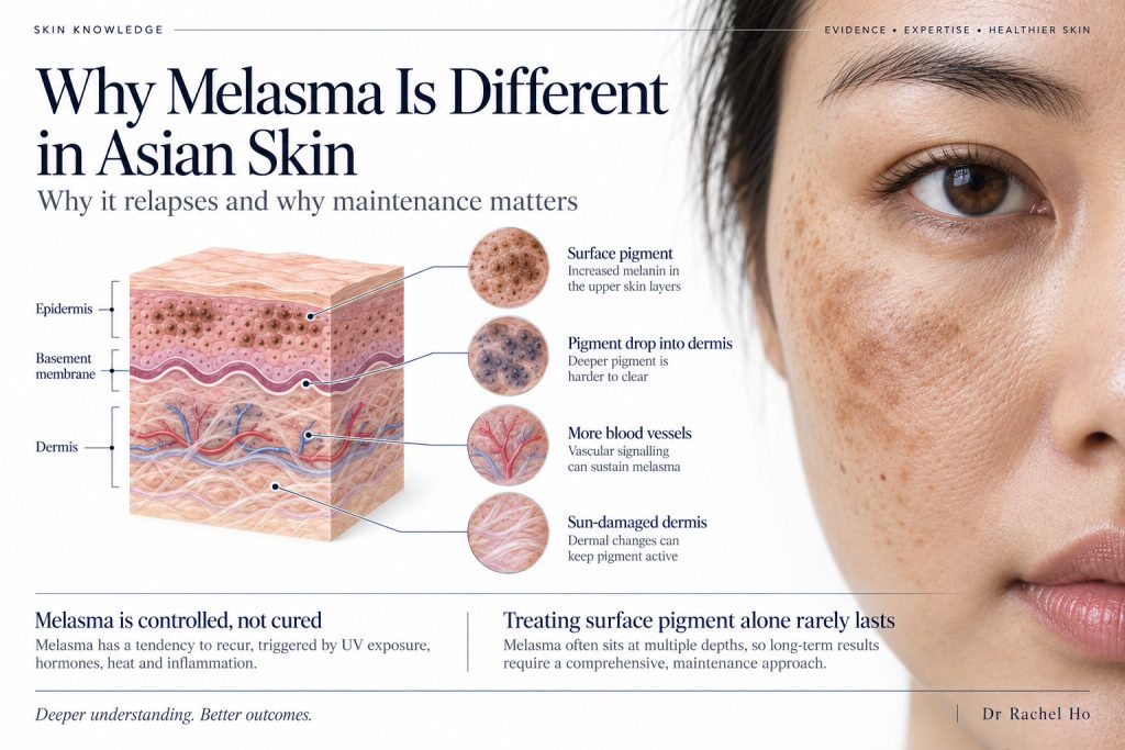
Melasma involves pigment activity alongside changes in blood vessels, the basement membrane and the skin’s supporting tissue. These findings help explain why treatment considers the underlying skin changes as well as the visible brown patches.
What helps hyperpigmentation?
Effective hyperpigmentation treatment must address both the visible dark spots on your skin and the underlying cellular triggers that cause reactivation of dark spots1,10. In my practice, I always tailor the approach based my patients’ medical diagnosis, skin condition and response to past treatments.
A successful, long-term strategy typically involves these four pillars:
Photoprotection
Daily broad-spectrum sunscreen is the non-negotiable foundation of melasma and pigmentation care, especially with Singapore’s high UV index all year round. To limit continued pigment stimulation from visible light, I highly recommend tinted formulations containing iron oxides10,12. My clinic’s guide to sunscreen for pigmentation and melasma explains exactly how to choose the right sun protection for melasma in Asians.
Targeted skincare
Over-the-counter active ingredients can gradually lighten pigmentation by inhibiting melanin production, blocking pigment transfer, or accelerating cell turnover18. However, because skin inflammation can paradoxically worsen pigmentation, I would advise avoiding layering excessive actives and prioritising your skincare barrier before layering more products. My guide to skincare for pigmentation and dark spots breaks down these ingredients and my guide to the skin barrier will take you through how to protect your skin while using strong actives.
Prescription medications
For stubborn conditions like melasma, medical intervention has a strongly established role. This includes doctor-supervised hydroquinone combination creams or oral tranexamic acid, which can be considered following a thorough medical assessment10,18. My article on tranexamic acid for melasma discusses the different formulations and medical considerations.
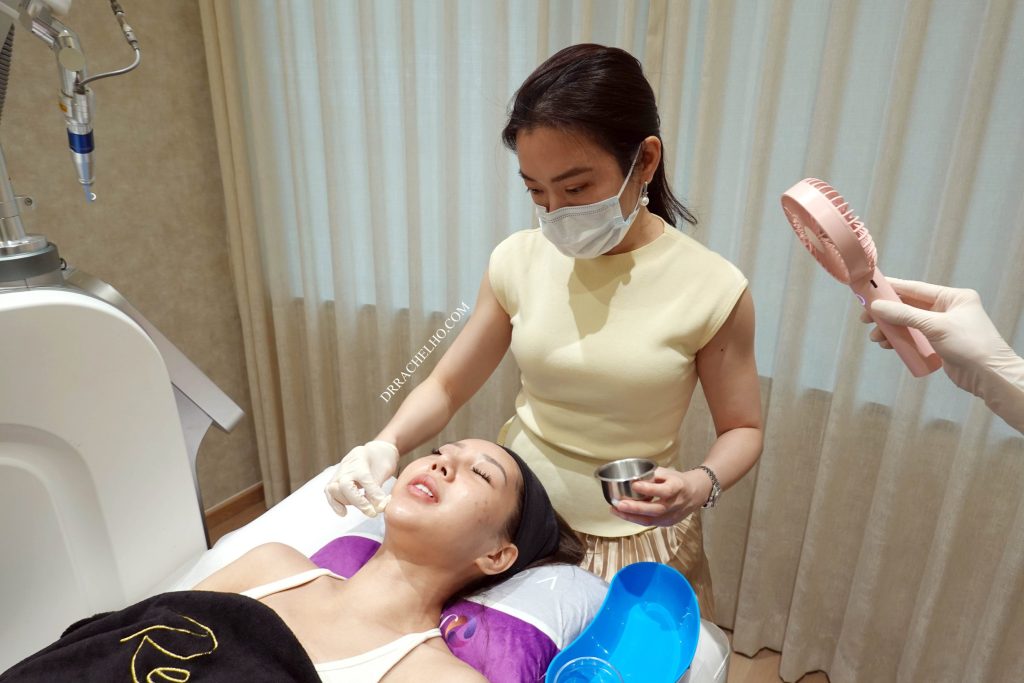
Administering a chemical peel for my patient in The Skin Longevity Clinic.
In-clinic procedures
Chemical peels, pico laser and RF microneedling treatments can precisely target pigment deposits and complement your topical routine1,10 . For melasma specifically, chemical peels are an excellent supplement to medical treatment, while pico lasers are generally introduced when initial therapies have provided an incomplete response. My pico laser review explains the clinical evidence and why the same laser yields very different results depending on the specific pigmentation condition.
Why melasma is different in Asian skin
Although melasma is regarded as a type of hyperpigmentation, patients with melasma actually have more than excess melanin in the skin. Histological studies of melasma have identified solar elastosis, disruption of the basement membrane, increased blood vessels and mast cells, and pigment within the dermis3,4. A 2026 review proposes that a photoaged dermal environment, including senescent fibroblasts and vascular signalling, helps sustain melanocyte activity6.
These findings explains two things my patients with melasma notice:
- Melasma recurs after it clears, because the dermis keeps signalling.
- Treatments aimed only at epidermal pigmentation clearance give short-lived results.
The complexity and relapsing nature of melasma means that the aim of treatments is to control the hyperpigmentation and its recurrence and not to cure melasma.
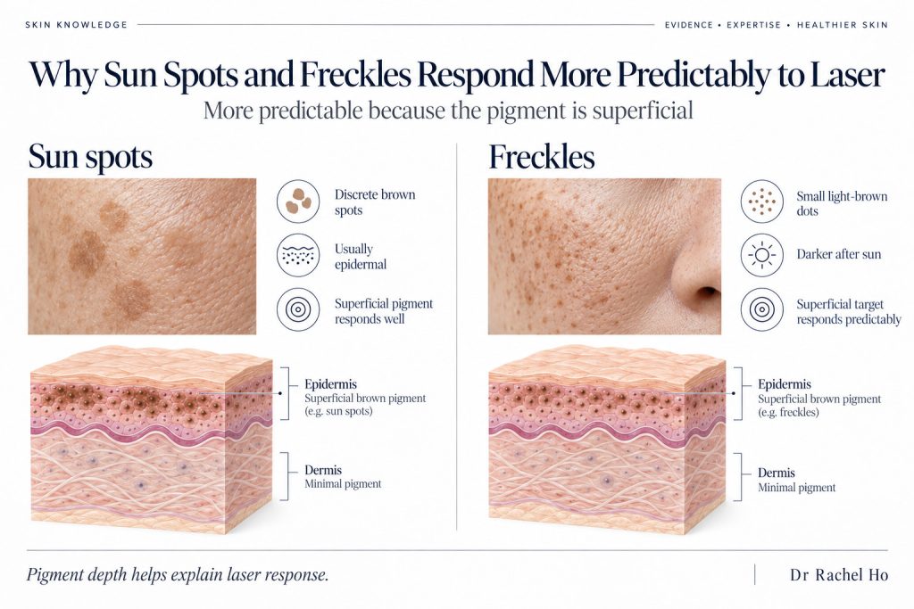
Sun spots or solar lentigines are associated with accumulated sun exposure, while freckles reflect an inherited tendency and become darker with sunlight. Both typically involve pigmentation in the epidermis, the outer skin layer illustrated in this comparison.
Why do solar lentigines and freckles respond faster to laser treatments?
Solar lentigines and freckles are more predictable pigmentary conditions to treat, partly because the pigment is located in the epidermis.
- Sun spots/ solar lentigines: In a study of 43 solar lentigines in 20 Asian women with skin types III or IV, a 532 nm picosecond laser achieved more than 75% clearance in 40 lesions, or approximately 93%, after one treatment [20]. PIH occurred in 4.65% of treated lesions20. PIH occurred in under 5% of spots20.
- Freckles: In a randomised split-face trial in Asian patients, Q-switched laser cleared freckles better than intense pulsed light21.
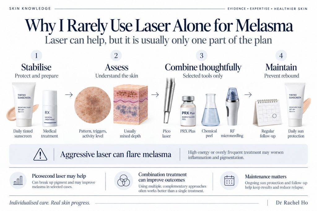
A summary of my clinical approach to melasma care, combining assessment, sun protection, medical treatment and maintenance. Pico laser, chemical peels, PRX Plus and radiofrequency microneedling appear as options discussed in the article, rather than a standard combination for every patient.
Why I rarely use lasers as a standalone treatment for melasma
Lasers, especially pico lasers, can help lighten melasma7. However, given the deeper skin involvement with melasma, and the reactive nature of melasma, I find that combining different modalities of pigmentation treatment gives a synergistic and safer approach. In my clinic, The Skin Longevity Clinic, some of these modalities include chemical peels for exfoliation of superficial hyperpigmentation, PRX Plus to suppress melanin formation and radiofrequency microneedling to disrupt melanin clusters and reinforce the basement membrane to reduce pigment incontinence.
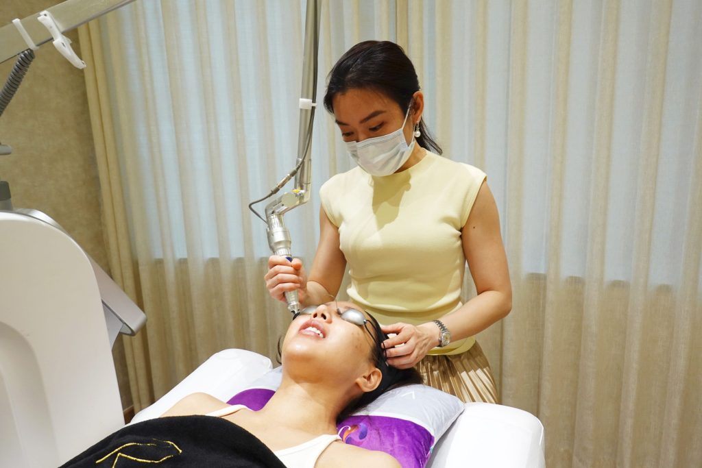
Administering pico laser for my patient in The Skin Longevity Clinic
What studies on pico laser studies show for melasma
A review of six randomised trials found improvement in melasma severity with 1064 nm picosecond treatment, although results varied between protocols7. The 755 nm pico laser studies showed no statistically significant advantage over topical agents, and PIH occurred in some studies7.
In a separate trial involving 18 participants, picosecond and nanosecond 1064 nm lasers produced similar improvement on opposite sides of the face8. The picosecond treatment was less painful, but recurrence was reported with both approaches8. For a more in-depth read on pico laser, please see my pico laser review.
Studies on multi-modality approaches to melasma treatments
The 2026 international melasma consensus places photoprotection and supervised topical treatment at the foundation of care, with laser considered for refractory melasma10. A retrospective study comparing 56 patients receiving laser toning with RF microneedling against 58 receiving toning alone found greater improvement with the combination11.
3 factors that have changed my practice: tinted sunscreens, tranexamic acid and RF microneedling
Visible light protection with tinted sunscreen
Besides UV light, high energy visible light from the sun is recognised as a trigger for melasma. A tinted sunscreen containing suitable iron oxides can add visible light protection to ultraviolet protection, which is relevant to melasma12. In a randomised trial involving 68 people, ultraviolet plus visible light protection produced greater improvement than ultraviolet protection alone when both groups also used hydroquinone12. For my sunscreen recommendations, please read my Sunscreen Reviews 2025.
Tranexamic acid
Tranexamic acid is an antifibrinolytic medicine that reduces the breakdown of blood clots15. Oral tranexamic acid is used off label for selected patients with melasma after assessment of their medical history and clotting risk15. Tranexamic acid works by inhibiting the plasmin-keratinocyte-plasminogen system, reducing α-MSH-driven melanogenesis, and also decreasing mast cells and angiogenesis relevant to melasma.
A randomised trial enrolled 44 patients with moderate to severe melasma and compared oral tranexamic acid with placebo alongside sunscreen14. At three months, the modified Melasma Area and Severity Index fell by 49% with tranexamic acid and 18% with placebo14.
Due to the risk of blood clotting due to oral tranexamic acid, I selectively prescribe tranexamic acid to my patients. Instead, I prefer intradermal delivery of tranexamic acid into the skin via radiofrequency microneedling and Local Dynamic Micromassage ultrasound treatment in my clinic to bypass the systemic side effects of oral tranexamic acid.
I have also previously shared more about the skincare science of tranexamic skincare in my guide to tranexamic acid.
Radiofrequency (RF) microneedling for melasma
RF microneedling delivers radiofrequency energy through fine needles to create controlled heating within the skin16. Histological studies included in a 2026 review reported changes in dermal senescence markers and partial restoration of basement membrane integrity with RF microneedling, although devices and protocols varied in the studies16.
A 2026 systematic review of 12 studies of RF microneedling for melasma reported improvements in melasma severity, with some benefits measured up to six months16. A separate retrospective study of 50 patients reported a median individual reduction in melasma severity of 32.3%17.
Combining tranexamic acid with RF microneedling has been shown to also enhance dermal penetration of tranexamic acid19. A split-face trial found microneedling with topical 0.4% tranexamic acid achieved comparable “good-to-excellent” outcomes (70%) to TCA 20% chemical peels19.
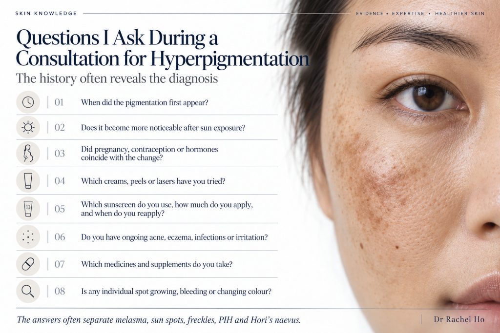
A hyperpigmentation consultation considers the timeline of hyperpigmentation developments, possible triggers, previous treatments, sunscreen habits and medical history. These questions help shape my treatment recommendations and identify changes that need closer examination.
Questions I ask during a consultation for hyperpigmentation
Besides identifying the nature of hyperpigmentation, I find it necessary to understand predisposing factors in my patients’ lifestyle and medical history that can influence their recovery, results and recurrences. This information helps to shape recommendations for treatments and to stage them safely. These are some of the questions that I ask during a typical consultation for hyperpigmentation:
- When did the pigmentation first appear, and how has it changed?
- Does it become more noticeable after sun exposure?
- Did pregnancy, contraception or other hormonal treatment coincide with the changes?
- Which creams, peels or lasers have you tried, and how did your skin react?
- Which sunscreen do you use, how much do you apply and when do you reapply?
- Do you have ongoing acne, eczema, infections or irritation?
- Which medicines and supplements do you take?
- Is any individual spot growing, bleeding or changing colour?
More about hyperpigmentation treatment in Singapore
My clinic’s guide to pigmentation treatment in Singapore covers treatment options, procedure details and other considerations. Besides in-clinic treatments, skincare for lightening hyperpigmentation has been discussed in How to Lighten Pigmentation and Dark Spots with Skin Care.
Frequently asked questions about hyperpigmentation
Can melasma be cured?
Melasma can improve substantially, but the recurrences can occur even after successful treatment4. Sunscreen and an appropriate maintenance plan help manage that tendency10. I would say that the goal of melasma treatment is control, not cure.
Why did my pigmentation get darker after laser treatments?
Inflammation following a procedure can cause PIH, while melasma can also recur after improvement5,8. The timing, distribution and baseline condition of the skin before treatment guide the differential diagnosis1.
Is it melasma or sun spots?
Melasma usually forms wider, symmetrical patches with ill defined edges, while solar lentigines are more discrete brown spots2,4. Their histories also differ, including hormonal influences in melasma and accumulated sunlight in solar lentigines2,4.
Do creams remove Hori’s naevus?
Hori’s naevus involves deeper deposition of melanin in the dermis and topical lightening creams generally have limited benefits for this pigmentation1. Pigment lasers are commonly considered for treatment of Hori’s naevus1. For a comprehensive guide to understanding how eye creams work and how to choose one for your under eye concerns, please refer to Do Eye Creams Work for Dark Eye Circles?
Does sunscreen really matter indoors?
Given Singapore’s high UV index all year around, I would recommend wearing broad spectrum sunscreen in the daytime even if you are indoors. For patients with melasma I would recommend tinted sunscreen to add visible light protection to ultraviolet protection12,13.
About Dr Rachel Ho
Dr Rachel Ho is the founder of The Skin Longevity Clinic in Singapore. She holds an MBBS from the National University of Singapore, a Master of Medicine and MRCS, and has 16 years of clinical experience. She is currently pursuing her Graduate Certificate in Healthy Longevity Medicine through the NUS Academy for Healthy Longevity at the Yong Loo Lin School of Medicine till the end of 2026. She writes about skin science, skincare ingredients and evidence-based aesthetic medicine on drrachelho.com.
References
- Passeron T, Genedy R, Salah L, et al. Laser treatment of hyperpigmented lesions: position statement of the European Society of Laser in Dermatology. Journal of the European Academy of Dermatology and Venereology. 2019;33(6):987–1005. DOI: 10.1111/jdv.15497. PMID: 30873649.
- Nautiyal A, Wairkar S. Management of hyperpigmentation: Current treatments and emerging therapies. Pigment Cell & Melanoma Research. 2021;34(6):1000–1014. DOI: 10.1111/pcmr.12986. PMID: 33998768.
- Phansuk K, Vachiramon V, Jurairattanaporn N, Chanprapaph K, Rattananukrom T. Dermal Pathology in Melasma: An Update Review. Clinical, Cosmetic and Investigational Dermatology. 2022;15:11–19. DOI: 10.2147/CCID.S343332. PMID: 35023942.
- Espósito ACC, et al. Update on Melasma—Part I: Pathogenesis. Dermatology and Therapy. 2022;12(9):1967–1988. DOI: 10.1007/s13555-022-00779-x. PMID: 35904706.
- Chaowattanapanit S, Silpa-Archa N, Kohli I, Lim HW, Hamzavi I. Postinflammatory hyperpigmentation: A comprehensive overview: Treatment options and prevention. Journal of the American Academy of Dermatology. 2017;77(4):607–621. DOI: 10.1016/j.jaad.2017.01.036. PMID: 28917452.
- Kang HY, Picardo M. Melasma: When Dermal Photoageing Precedes Melanocyte Ageing. Experimental Dermatology. 2026;35(9):e70364. DOI: 10.1111/exd.70364. PMID: 42746716.
- Feng J, Shen S, Song X, Xiang W. Efficacy and safety of picosecond laser for the treatment of melasma: a systematic review and meta-analysis. Lasers in Medical Science. 2023;38(1):84. DOI: 10.1007/s10103-023-03744-y. PMID: 36897459.
- Feng J, Huang L. Comparison of Picosecond and Nanosecond Nd:YAG 1064-nm Lasers in the Treatment of Melasma: A Split-Face Randomized Clinical Trial. Plastic and Reconstructive Surgery. 2023;151(4):772–777. DOI: 10.1097/PRS.0000000000009994. PMID: 36729879.
- Chan NPY, Ho SGY, Shek SYN, Yeung CK, Chan HH. A case series of facial depigmentation associated with low fluence Q-switched 1,064 nm Nd:YAG laser for skin rejuvenation and melasma. Lasers in Surgery and Medicine. 2010;42(8):712–719. DOI: 10.1002/lsm.20956. PMID: 20848553.
- Sarkar R, Desai SR, Sinha S, et al. Delphi consensus on melasma management by international experts and pigmentary disorders society. Journal of the European Academy of Dermatology and Venereology. 2026;40(4):680–692. DOI: 10.1111/jdv.70066. PMID: 40996222.
- Kwon HH, Choi SC, Jung JY, Park GH. Combined treatment of melasma involving low-fluence Q-switched Nd:YAG laser and fractional microneedling radiofrequency. Journal of Dermatological Treatment. 2019;30(4):352–356. DOI: 10.1080/09546634.2018.1516858. PMID: 30160539.
- Castanedo-Cazares JP, Hernandez-Blanco D, Carlos-Ortega B, Fuentes-Ahumada C, Torres-Álvarez B. Near-visible light and UV photoprotection in the treatment of melasma: a double-blind randomized trial. Photodermatology, Photoimmunology & Photomedicine. 2014;30(1):35–42. DOI: 10.1111/phpp.12086. PMID: 24313385.
- Polena H, Queille-Roussel C, Graizeau C, Duteil L, Sayag M, Passeron T. Comparison of Visible Light-Protective Tinted Sunscreen to Untinted Sunscreen to Protect Melasma Patients During Summer: A Prospective Randomized Investigator-Blinded Study. Journal of Cosmetic Dermatology. 2025;24(10):e70450. DOI: 10.1111/jocd.70450. PMID: 41014037.
- Del Rosario E, Florez-Pollack S, Zapata L Jr, et al. Randomized, placebo-controlled, double-blind study of oral tranexamic acid in the treatment of moderate-to-severe melasma. Journal of the American Academy of Dermatology. 2018;78(2):363–369. DOI: 10.1016/j.jaad.2017.09.053. PMID: 28987494.
- Godse K, Sarkar R, Mysore V, et al. Oral Tranexamic Acid for the Treatment of Melasma: Evidence and Experience-Based Consensus Statement from Indian Experts. Indian Journal of Dermatology. 2023;68(2):178–185. DOI: 10.4103/ijd.ijd_266_22. PMID: 37275826.
- Kumar N, Lin S, Luthra A, Patel T, Bhatia AC. The Efficacy and Safety of Radiofrequency Microneedling for Melasma: A Systematic Review and Qualitative Evidence Synthesis. Aesthetic Surgery Journal Open Forum. 2026;8:ojag075. DOI: 10.1093/asjof/ojag075. PMID: 42253423. OUP Academic
- Kim HB, Lee SY, Um JY, Baek SY, Kim HO, Kumar N. Clinical Effects and Safety of Radiofrequency Microneedling for the Management of Melasma: A Retrospective Study. Aesthetic Surgery Journal Open Forum. 2026;8:ojag143. DOI: 10.1093/asjof/ojag143. PMID: 42582527.
- Gan C, Rodrigues M. An Update on New and Existing Treatments for the Management of Melasma. American Journal of Clinical Dermatology. 2024;25(5):717–733. DOI: 10.1007/s40257-024-00863-2. PMID: 38896402.
- Sayed DS, Fathy E, Mahmoud SM, Hassan H, Tawfik YM. TCA 20% peeling versus either cryopeeling or topical tranexamic acid combined with microneedling in treatment of melasma: a split face randomized clinical trial. Archives of Dermatological Research. 2025;317:848. DOI: 10.1007/s00403-025-04352-2. Negishi K, Akita H, Matsunaga Y. Prospective study of removing solar lentigines in Asians using a novel dual-wavelength and dual-pulse width picosecond laser. Lasers in Surgery and Medicine. 2018;50(8):851–858. DOI: 10.1002/lsm.22820. PMID: 29608215.
- Wang CC, Sue YM, Yang CH, Chen CK. A comparison of Q-switched alexandrite laser and intense pulsed light for the treatment of freckles and lentigines in Asian persons: a randomized, physician-blinded, split-face comparative trial. Journal of the American Academy of Dermatology. 2006;54(5):804–810. DOI: 10.1016/j.jaad.2006.01.012. PMID: 16635661.

No Comments